
ChromaTwist Dye to Antibody Conjugation and Multicolour Flow Cytometric Evaluation
Executive Summary
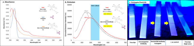
- The ChromaTwist dye (Gen-2-Dye) has been conjugated to the CD8 antibody (Ab) as shown by UV/Vis (Figure 1a) and emission spectroscopy (Figure 1b). In addition, the conjugate was shown to be active and bound to Protein A and G in the Abcam Conjugation Check kit (Figure 1c).
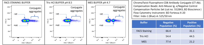
- The Gen-2-DyeCD8 conjugate when incubated with Anti-Mouse Ig, κ/Negative Control Compensation Particles Set in three different buffers lead to positive particle staining (BD Fortessa X-20 Flow Cytometer) (Figure 2) ranging from 21.2-44.5%. BUV395●CD8 was used as a commercial comparison in FACS staining buffer and gave 38.7 % +ve population compared to 31.1% for the Gen-2-Dye●CD8 conjugate. More technical details are set out below.
ChromaTwist Dye to CD8 Antibody Conjugation
A ChromaTwist dye (Gen-2-Dye) has been conjugated to CD8 RPA-T8 antibody (Ab). Characterisation of the Gen-2-Dye●CD8 conjugate (post Pierce™ Dye Removal Column and Amicon Ultra-0.5 (30 kDa) centrifugal filtration) to rigorously exclude free dye) with absorbance (Figure 1a) and emission (Figure 1b) spectroscopies together with a Conjugate Check Kit (Figure 2c) provided evidence of successful conjugation, and removal of free dye. Degree of labelling of upto 10 CT dyes per Ab was achieved.
Flow Cytometric Evaluation of the CT-Ab Conjugate
The headline result is that postive staining was achieved using the CT dye-Ab conjugate with Anti-Mouse Ig, κ/Negative Control Compensation Particles Set (Figure 2). This staining was observed using the Indo-1 (blue) filter, which only detects part of the tail of the Gen-2-Dye●CD8 conjugate’s fluorescence (Figure 1b)). The staining revealed a buffer dependence with MES buffer affording a 21.2% positive stain, which increased with FACS (31.1%) and Tris HCl (44.5%) buffer. The BUV395●CD8 conjugate was used as a commercial comparison with FACS staining buffer and gave a positive stain population of 38.7%, relative to 31.1% with the Gen-2-Dye●CD8 conjugate. Conjugate aggregates were observed at room temp which inhibited partcle binding. Therefore, to reduce this aggregation bead staining was carried out at 37 °C, which reduced aggregate formation and enable particle binding, as shown in Figure 2.
Read more about ChromaTwist dye development for Spectral Flow Cytometry.