
Background: ChromaTwist Dyes for Flow Cytometry: The chemical modularity of ChromaTwist fluorescent dye technology has allowed 50+ UV excitable fluorescent dyes to be created that emit across the visible spectrum (395 – 635 nm, and potential for reaching into IR emission), without the need for tandem dye based approaches, and their drawbacks for end-users. Previously we demonstrated that the ChromaTwist blue dye-antibody (CD4 & CD8) conjugates have utility in both multicolour and spectral flow cytometry (BD Bioscience Fortessa X-20, Cytek Aurora (355 nm); Sony Biotechnology ID7000 (320 nm, 355 nm), ThermoFisher BigFoot (355 nm)).
Potential of the ChromaTwist UV Excitable Dye Technology in Flow Cytometry:
Multicolour Flow Cytometry: The chemical tunability of the ChromaTwist dyes means that we are able to match the fluorescent emission outputs to the 6 visible flow cytometry filters and ultimately the 2 IR filters, and bring to the multicolour market a portfolio of UV excitable molecular based dyes, without the end-user drawbacks associated with tandem polymer dyes.
UV Spectral Flow Cytometry: The chemical tunability of the ChromaTwist dyes means that we will be able to go far beyond the multicolour 8 dye range and be able to present a portfolio of 50+ UV excitable dyes (320 nm and/or 355 nm), each with a unique spectral fingerprint. Ultimately this means ChromaTwist could offer 50+ ChromaTwist-dye antibody conjugates which are all excited by 320 and/or 355 nm UV laser.
 Conclusion: Significant improvement of staining indices since Technical Note 5 (May 2023) and the conjugates are photostable over a period of at least a week (stored in the refrigerator 4 °C; stability testing is on-going).
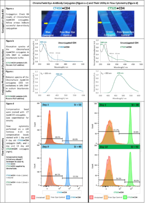
CT Dye●CD4 Conjugate Detection using a Conjugate Check Kit, Figure a: Successful conjugation is evident from the fluorescence observed at the “test line” upon irradiation with 302 nm light, indicating that the CD4 antibody was indeed labelled with the ChromaTwist dyes. The free dye controls (samples without antibody) show no staining at the test line.
Comparison of CT Dye●CD4 Conjugate Absorption Spectra, Figure b: The absorption spectra of CT450●CD4 and CT525●CD4 are compared to the unconjugated antibody, revealing the conjugate has an absorption profile containing elements of the dye and antibody, and a significant shift in the antibody λmax (20 nm) giving good evidence of conjugation. The absorption spectra of conjugates show that they are excited by the conventional 355 nm laser used in flow cytometry, but also the 320 nm laser system recently developed by Sony Biotechnology in their ID7000 instrument.
CT Dye●CD4 Conjugate Emission Spectra, Figure c: The emission spectra of CT450●CD4 and CT525●CD4 conjugates show peak emission is observed at 465 nm and 479 nm respectively, which is congruent with the free blue and 525 emission profiles.
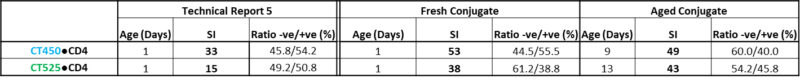
UltraComp eBeads™ Staining with CT Dye●CD4 Conjugate and Flow Cytometry, Figure d: Stained UltraComp eBeads™ were analysed on a LSR Fortessa X-20. Beads were gated to exclude debris and doublets and fluorescence is observed in the indo-1 (violet) and indo-1 (blue) fluorescence channels for the 1 day old blue and green CT450●CD4 and CT525●CD4 conjugates, respectively, affording staining indices of 53 and 38. The CT450●CD4 and CT525●CD4 conjugates were tested again after 9 and 13 days respectively, affording staining indices of 49 and 43, indicating stability over at least a week in (on-going) stability tests.
ChromaTwist is currently working on a development program to improve dye conjugate purification, optimise Degree of Labelling (dye/protein ratio) and perform cell staining. We are also developing technology to push these staining indices up by factors of 2, 3, 4, 6, 8, and 9 and potentially beyond via a scalable amplification technology and expand our conjugated dye portfolio to include red emissive dyes.
Conjugate Check Kit Experimental:
Solutions of the CT dye●CD4 conjugates (5 μL of conjugate, 35 μL 1x running buffer/1 % BSA) were run on a Conjugation Check Kit (Abcam ab236554).
Staining Beads and Flow Cytometry Experimental:
- Vortex (20 s) and add compensation beads (Invitrogen 01-2222-42, 25 µL) to a 1.5 mL Eppendorf.
- Add ChromaTwist dye●CD4 antibody conjugate (0.5 µL) to the beads, ensuring the conjugate is deposited directly in to the beads.
- Incubate the sample in the dark (30 min, 21 °C).
- Add staining buffer (Biolegend cat no. 420201, 1400 µL) and vortex (3 s).
- Centrifuge to wash (600 g, 5 min).
- Remove supernatant, leaving behind the bead pellet.
- Resuspend the bead pellet in staining buffer (1400 µl), vortex (3 s) and centrifuge (600 g, 5 min) – repeat steps ⑥ and ⑦ for a total of three washes.
- Resuspend the bead pellet in 500 µL staining buffer, vortex (3 s) and transfer the sample to a FACS tube.
- Samples are analysed on a LSR Fortessa X-20 flow cytometer. Beads are gated to exclude debris and doublets and fluorescence is observed in the indo-1 (Violet) ≡ 450/50 and indo-1 (Blue) ≡ 525/50 fluorescence channels for the CT450●CD4 and CT525●CD4 conjugates, respectively.